Cancer Metabolism and Signaling in the Tumor Microenvironment
From metabolic reprogramming in cancer cells to creating nucleotide imbalances. These experts are advancing the field of medical research and cancer treatment.
Published August 6, 2024
By Megan Prescott, PhD
Program Manager for Life Sciences
What causes a normal cell to become a cancer cell? How do cancer cells cooperate to form a tumor? How can we interrupt these processes to inhibit cancer growth? Can nutrients directly modulate disease progression and therapeutic response?
These and related questions were the focus of a conference held on April 17, 2024. The conference was presented by The New York Academy of Sciences and NYU Langone Health. The program held at the NYU Medical Center, included presentations by world renowned researchers in the field of cancer metabolism. The goal was to understand how these findings can be translated into therapies that will impact the lives of patients.
Metabolic pathways represent a powerful, yet underappreciated set of therapeutic targets for cancer. They play a crucial role in tumorigenesis, the transformation of normal cells into cancerous ones. Oncogenic mutations may alter these metabolic pathways, enabling cells to extract energy from their surroundings. Additionally, they manipulate signaling pathways to drive tumor development and advancement.
Mitochondrial Adaptations and Signaling in Tumors

Photo by Nick Fetty/The New York Academy of Sciences
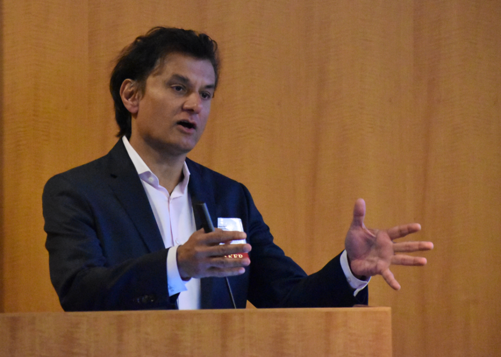
Opening speaker, Navdeep Chandel, PhD, David W. Cugell, MD Professor at Northwestern University, described how metabolic reprogramming in cancer cells is directly triggered by oncogenes. Some of the metabolic genes important for oncogenesis include those found in the electron transport chain (ETC) of mitochondria.
Since mitochondria are a biosynthetic and bioenergetic hub inside of cells, many types of cancer cells, which proliferate quickly and have high energy demands, rely heavily on mitochondria for their survival. Electron transport chain function is responsible for providing metabolites linked to the tricarboxylic acid cycle (TCA). This provides the building blocks for cell proliferation. Dr. Chandel has shown that the widely used anti-diabetic drug metformin has anti-tumor effects through inhibition of Mitochondrial Complex I of the ETC within cancer cells.
Immune-dependent attenuation of tumor growth was seen in work from Pere Puigserver, PhD. Dr. Puigserver is a professor of cell biology at Harvard Medical School and the Dana-Farber Cancer Institute. Mitochondrial Complex I inhibition in tumors triggered by deletion of the subunit Ndufs4, increases the activation status of CD8+ T Cells and Natural Killer cells within the tumor environment. This finding has potential implications in the field of immunotherapy.
Oxygen, Iron, and Vitamins in the Tumor Microenvironment
Electron Transfer Reactions in the mitochondria are facilitated by iron-sulfur containing proteins. Isha Jain, PhD, assistant professor in biochemistry and biophysics in the School of Medicine at the University of California, San Francisco, showed how these proteins are damaged in high oxygen (hyperoxic) conditions. While researchers have studied the detrimental effects of low oxygen on the body for a long time, Dr. Jain’s work focuses on discovering why too much oxygen is toxic in some cases.
“We found that certain proteins that contain iron, basically rust in high oxygen, and that’s why things go wrong,” she explained. Her work opens the question of whether treatments that can be developed to protect or repair these proteins.

Photo by Nick Fetty/The New York Academy of Sciences
Research from Richard Possemato, PhD, associate professor in pathology at the NYU Grossman School of Medicine, showed that iron-sulfur clusters are important for tumor growth in breast cancer. DNA Polymerase Epsilon (POLE) contains an iron-sulfur cluster, and inhibition of POLE by disrupting its iron-sulfur cluster eradicates tumors in a mouse model of triple negative breast cancer. Furthermore, tumor eradication by this method induces adaptive immunity, and researchers were unable to grow tumors in these mice again.
Recent work has emphasized that the stressful conditions of the tumor microenvironment. Parts of the tumor periodically experience limited availability of primary nutrients and oxygen. This also affects the metabolism of cancer cells. Cell proliferation, the hallmark of cancer, is metabolically demanding. It requires energy and cellular ‘building blocks’ in the form of amino acids for proteins, fatty acids for lipids, and nucleotides for DNA and RNA.
How Cells Rewire Their Metabolism
Gerta Hoxhaj, PhD, assistant professor in the Children’s Medical Center Research Institute at the University of Texas Southwestern Medical Center, described how cells rewire their metabolism to fuel the growth and survival of cancer cells. Cells need a constant supply of nucleotides to grow, proliferate, and function.
Cells can either get their supply of purine nucleotides from simple molecules like amino acids by de novo synthesis or can recover purines from the breakdown of DNA and RNA through the salvage pathway. While de novo synthesis and salvage pathways contribute similarly to purine pools in tumors, the salvage pathway is critical for tumor growth in mouse models of liver cancer, among others.
Research from Celeste Simon, PhD, the Arthur H. Rubenstein, MBBCh Professor at the University of Pennsylvania, demonstrates that metabolic crosstalk is also important in Pancreatic Ductal Adenocarcinoma (PDAC), the second leading cause of cancer related death in 2023. Fibroblasts help PDAC cells survive by supplying these tumor cells with unsaturated fatty acids for the maintenance of lipid homeostasis in low oxygen (hypoxic) and nutrient-poor environments. Finding drugs to disrupt this cross-talk could be a novel metabolic target in PDAC treatment.
Cancer Cell Intrinsic and Extrinsic Determinants of Tumor Metabolism
The tumor microenvironment of PDAC has abundant fibroblasts of different lineages and functions according to Mara Sherman, PhD, head of the Mara Sherman lab at Memorial Sloan Kettering Cancer Center. “We identified one lineage that promotes pancreatic cancer metastasis and seems to do so along nerves,” she said.

Social interactions between cancer cells, such as competition and cooperation, is an interest of Carlos Carmona-Fontaine, PhD, associate professor of biology at NYU. “The key currency for this cell-cell interaction is nutrients and other metabolites including oxygen,” he noted. Specifically, his presentation asked how amino acids become cooperative goods in low oxygen environments.
Amino acid starved cells cooperate to digest extracellular peptides: Both low, and high density, populations die without glutamine, but high-density populations recover when it is added back. The essential enzyme in this process is CNDP2. Inhibition of this form of cooperation impaired tumor growth.
The Impact of Blocking Adenosine Uptake in T-cells
Matthew Vander Heiden, PhD, Lester Wolfe Professor of Molecular Biology at MIT and director of the Koch Institute for Integrative Cancer Research, found that the nucleotide precursor adenosine suppresses anti-cancer immune responses. He presented work that showed blocking adenosine uptake in T-cells rescues proliferation and partially rescues cytokine production in these cells through salvaging pyrimidine nucleotides. Environmental conditions promoting nucleotide imbalance in T cells can regulate immune response, showing that if you can create nucleotide imbalances, then you can change cell fate.
This conference provided insight into metabolic changes, genes and pathways that support tumor growth and proliferation, and how this knowledge can inform new treatments that disrupt the strategies cancer cells depend on to survive.
